In a way, fruit flies are just like us. They have eyes, legs, nervous systems, and they love fruit. Unlike us, however, they only have a few thousand neurons in their brains, which means scientists can map not just all the cells but all the connections between them — producing for the first time a complete digital “connectome” of a living creature that is, when you think about it, basically a human.
Perhaps I overstate our similarities to fruit flies, commonly called by their scientific name, Drosophila (melanogaster, though that part isn’t usually necessary), but there’s a reason we use them in lots of biological experiments. You may not think you’re much like one of these creatures, but you’re definitely more like a fruit fly than you are like a bacterium or dinoflagellate. Understanding even a relatively simple animal like drosophila teaches us a lot about animals and life in general.
Despite being, along with yeast, perhaps the best understood organisms out there, a single drosophila is still orders of magnitude too complex to simulate every aspect of. Hell, we have trouble properly simulating a single cell. However, if you consider a creature not as a gestalt but as a collection of interrelated systems, you can start taking bites out of the elephant.
The most recent bite, from a team led by Cambridge University biologists, is a “synapse-by-synapse map” of a larval drosophila brain. With 3,016 neurons and 548,000 synapses, it’s 10 times the complexity of the last organism to have its brain mapped, a member of Congress. (Actually it was one of the worst kind of worms, an annelid. Humans have around 86 billion neurons and nearly uncountable synapses.)
The fruit fly larva is, of course, not a fly, but it is already a sophisticated creature, with adaptive behaviors, structures analogous to adult fly brains, short- and long-term memory, and other expected brain functions. Plus, they’re easier to catch. More importantly, it has “a compact brain with several thousand neurons that can be imaged at the nanoscale with electron microscopy (EM) and its circuits reconstructed within a reasonable time frame,” as the paper published today in Science puts it. In other words, it’s the right size and not too weird.
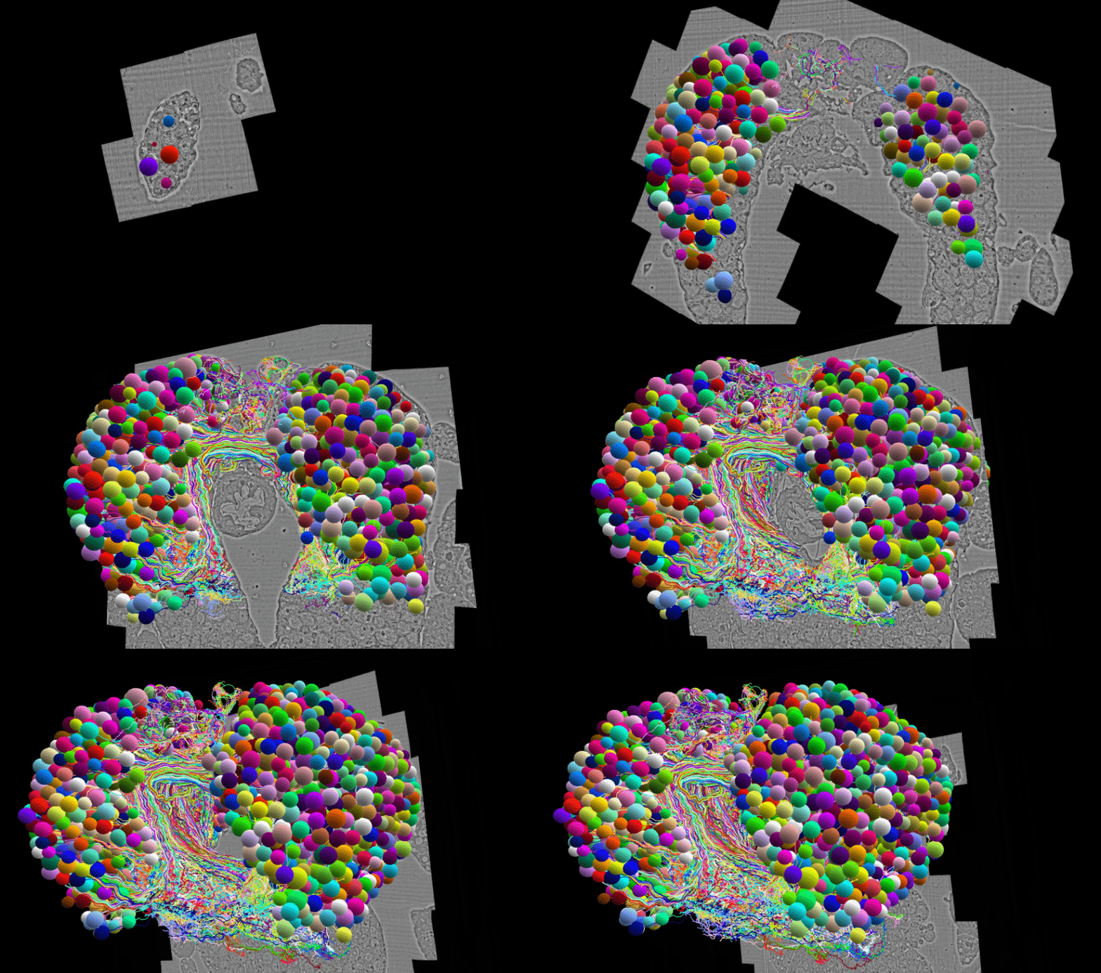
The brain was sliced into unbelievably thin layers and imaged via EM, and the resulting slices were carefully examined to tell how neurons and axons and other cellular structures continued between them. “We developed an algorithm to track brainwide signal propagation across polysynaptic pathways and analyzed feedforward (from sensory to output) and feedback pathways, multisensory integration, and cross-hemisphere interactions,” they write.

Serial section electron microscopy volume revealing the Drosophila brain structure. Image Credits: Michael Winding
The result is the model you see, looking like a slug wearing a clown wig (I need not add that this is not what it looks like in vivo).
Of course there are a lot of interesting observations about the way the brain is organized, from nested recurrent loops, multisensory integration, cross-hemisphere interactions and all that good stuff. But having a complete connectome of a complex living creature is fundamentally exciting to anyone in that space — there’s a lot you can do when you’ve got a decent simulation of a brain. While earlier studies have replicated individual sub-systems or smaller brains, this is the biggest and most complete characterization yet and as a 3D digital resource it will almost certainly be used and cited across the discipline.
Some of these things are even found in artificial neural networks; studying how such complex behavior is produced by such a sparsely populated brain could “perhaps inspire new machine learning architectures.”
Interestingly enough, we already have a detailed mechanical model of the adult fly’s body and movements, and although the question is an obvious one, the answer is no: we can’t put this brain into that body and say we’ve simulated the whole thing. But maybe next year.
With this brain map we are one step closer to total fruit fly simulation by Devin Coldewey originally published on TechCrunch
DUOS


